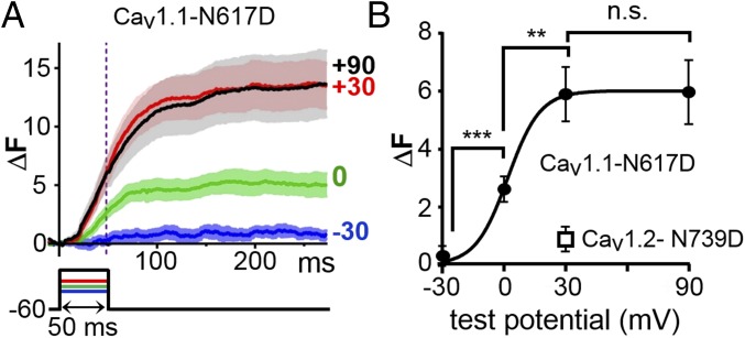
Fig. 5.
The voltage dependence of Ca2+ release in cells expressing five triad-junction proteins is similar to that of Ca2+ release in skeletal muscle. (A) RyR1-stable cells were transiently transfected with YFP–CaV1.1–N617D, β1a, Stac3–RFP, and JP2, loaded with Fluo-3 AM, and depolarized to the indicated potentials with the perforated patch technique. Data are presented as mean (solid line) ± SEM. Number of cells averaged was 10, 13, 24, and 22 for −30, 0, +30, and +90 mV, respectively, is shown. (B) Average ΔF, measured at 48 ms after the onset of depolarization (vertical dotted line in A), as a function of test potential. Based on Welch’s adjusted t test, the bracketed values were statistically different: ***P = 0.0006; **P = 0.002; n.s., not significantly different (P = 0.949). The data point for CaV1.2–N739D was obtained from the eight cells used to generate the average transient in Fig. 4D.

