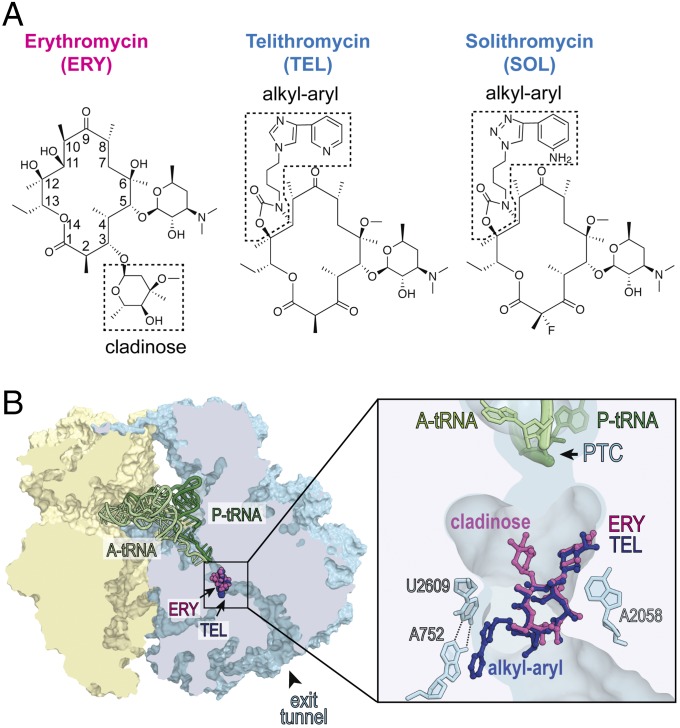
Fig. 1.
Bacteriostatic and bactericidal macrolide and ketolide antibiotics bind to the same site in the bacterial ribosome. (A) Chemical structures of bacteriostatic macrolide ERY and bactericidal ketolides TEL and SOL. The C3-cladinose sugar of ERY and the alkyl-aryl side chains in TEL and SOL are boxed. (B) Binding site of macrolides and ketolides in the ribosome. A cross section of the bacterial (Thermus thermophilus) 70S ribosome with ERY (purple) and TEL (blue) bound in the nascent peptide exit tunnel (PDB ID codes: 4V7X and 4V7Z, respectively, from ref. 11). Small subunit is yellow, large subunit is light blue, A-site tRNA is light green, and P-site tRNA is dark green. The zoomed-in image shows interactions of the C5 desosamine of both antibiotics with A2058 and of the alkyl-aryl side chain of TEL with the A752-U2609 base pair in the nascent peptide exit tunnel. The peptidyl-transferase center (PTC) is indicated.

