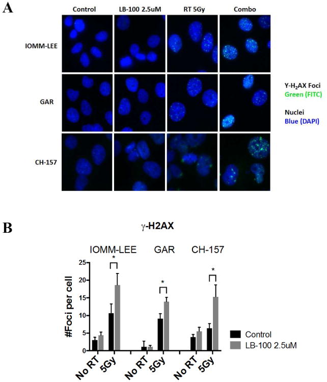
Figure 2.

LB-100 enhanced radiation induced DNA damage. (A) Immunofluorescence staining to detect γ-H2AX foci was performed in IOMM-LEE, GAR and CH-157 cells 48 hours after treatment. Cells were plated in 4-well chamber slides and allowed to attach (6-12 hours), and then treated with LB-100 (2.5 uM) for 3 hours prior to radiation (5 Gy). Foci were counted in at least 50 cells per experiment. Representative images for each cell line in each treatment group are shown. (B) Quantitative assessment of γ-H2AX foci per cell at 48 hours after radiation is shown. Data are the mean ± SEM from three independent experiments. *, P < 0.05 (student t-test comparing with or without LB-100 in the RT -treated groups.
