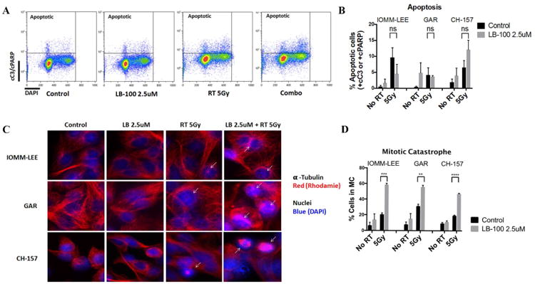
Figure 3.

LB-100 enhanced radiation induced mitotic catastrophe but not apoptosis. (A) Flow cytometry analysis was performed to determine rate of apoptosis in IOMM-LEE, GAR and CH-157 cells 48 hours after treatment. LB-100 was given 3 hours prior to RT. Double staining with cC3/cPARP-DAPI was used to label apopototic cells. Representative FACS plots were shown. (B). Quantification of the data showed no enhancement of apoptosis with LB-100 in RT treated groups (student t-test) (C) IOMM-LEE, GAR and CH-157 cells growing in chamber slides were exposed to LB 100 (2.5 umol/L) for 3 hours, irradiated (5 Gy), and 48 hours after radiation immunofluorescence staining was performed to assess the level of mitotic catastrophe. Nuclear fragmentation (defined as the presence of two or more distinct lobes within a single cell) was evaluated in at least 100 cells per treatment per experiment. Representative images for each cell line in each treatment group are shown. D, Quantitative assessment of percentage of cells in MC at 48 hours after radiation is shown. Data represents the mean ± SEM from three independent experiments. *, P < 0.05 (student t-test comparing with or without LB-100 in the RT -treated groups).
