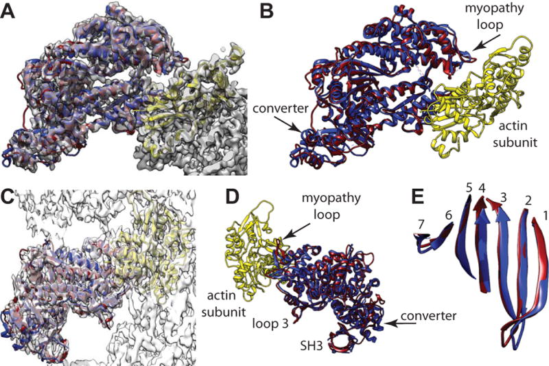
Figure 5.

Comparison of the actin bound, smMD and the nucleotide-free myosin-V MD crystal structure (PDB 1OE9), which has been aligned to the reconstruction using Chimera’s fitinmap utility. Coloring scheme has the actin subunit yellow, the smMD dark magenta, and the myosin-V MD blue. (A) View down the actin binding cleft showing the excellent fit of the myosin-V crystal structure even though not bound to actin. The myosin-V converter domain has a very similar position and orientation as the smMD converter. (B) Same view direction as panel A but with the map removed to shown the excellent alignment of the myosin-V helices with the corresponding smMD helices and loops. (C) View perpendicular to the helix axis showing the fit of the myosin-V coordinates within the acto-smMD reconstruction. (D) View from the opposite side without the map showing alignment of the myopathy loop, loop 3 and the SH3 domains. (E) View showing the excellent alignment of the 7-stranded transducer β-sheets.
