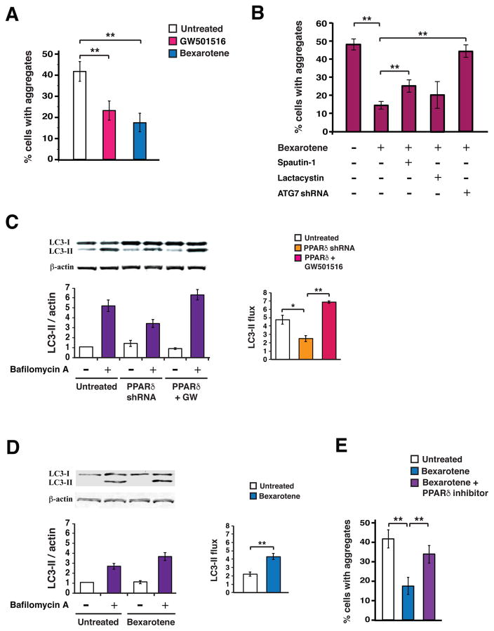
Figure 7. Bexarotene activation of PPARδ promotes proteostasis by inducing the autophagy pathway in mouse neurons.
(A) We quantified the percentage of Neuro2a cells containing htt protein aggregates, when transfected with a htt-104Q expression vector, treated for 24 hours with GW501516 (500 nM) or Bexarotene (1 μM), and exposed to H2O2 (25μM for 4 hours). **P < 0.01; ANOVA with post-hoc Tukey test. n = 30 – 50 cells / sample, 9 samples / condition. (B) We quantified the percentage of Neuro2a cells containing htt protein aggregates, when transfected with a htt-104Q expression vector, treated for 24 hours with Bexarotene (1 μM), Spautin-1 (10 nM) or Lactacystin (5 nM), and exposed to H2O2 (25μM for 4 hours). **P < 0.01; ANOVA with post-hoc Tukey test. n = 30 – 50 cells / sample, 9 – 12 samples / condition. (C) We performed microtubule-associated protein 1A/1B-light chain 3 (LC3) immunoblot analysis of Neuro2a cells cultured in normal media in the presence or absence of bafilomycin, and transfected with a PPARδ shRNA vector, or a PPARδ expression vector and treated with GW501516 (100 nM). β-actin served as a loading control. (D) We performed densitometry analysis of the LC3 immunoblotting results shown in (C) to determine autophagy flux. *P<0.05, **P < 0.01; ANOVA with post-hoc Tukey test. n = 3 independent experiments. (E) We quantified the percentage of Neuro2a cells containing htt protein aggregates, when transfected with a htt-104Q expression vector, treated with bexarotene (500 nM) alone, or bexarotene (500 nM) plus the PPARδ inhibitor GSK3787 (200 nM), and exposed to H2O2 (25μM for 4 hours). **P < 0.01; ANOVA with post-hoc Tukey test. n = 30 – 50 cells / sample, 9 samples / condition. Error bars = s.e.m.

