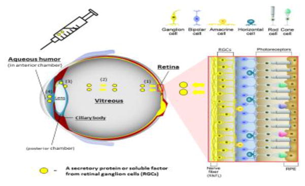
Figure 3 (Key figure). Structure of the neurosensory retina and access to molecular biomarkers of retinal ganglion cell health.
The retina is composed of various types of neurons (red box inset); photoreceptors (rods and cones) in the outer retina, intermediate neurons (bipolar, amacrine, and horizontal cells), and ganglion cells in the inner retina. Axons of retinal ganglion cell (RGC) come together to form the optic nerve that extends processes that synapse in the lateral geniculate nucleus, ultimately sending axons to the occipital (visual) cortex of the brain. Retinal pigmented epithelium (RPE), which is located external to the neurosensory retina, performs essential specialized functions such as phagocytosis of photoreceptor outer segments and recycling photopigments in the visual cycle.
Secreted proteins or soluble factors are released from RGCs into vitreous (1) and can diffuse throughout the vitreous body (2). In addition, they can be detected in the aqueous humor (AH) in the posterior chamber (3), and in the anterior chamber (4) of the eye. AH in anterior chamber is collected by a micro-invasive procedure called paracentesis through the peripheral cornea.

