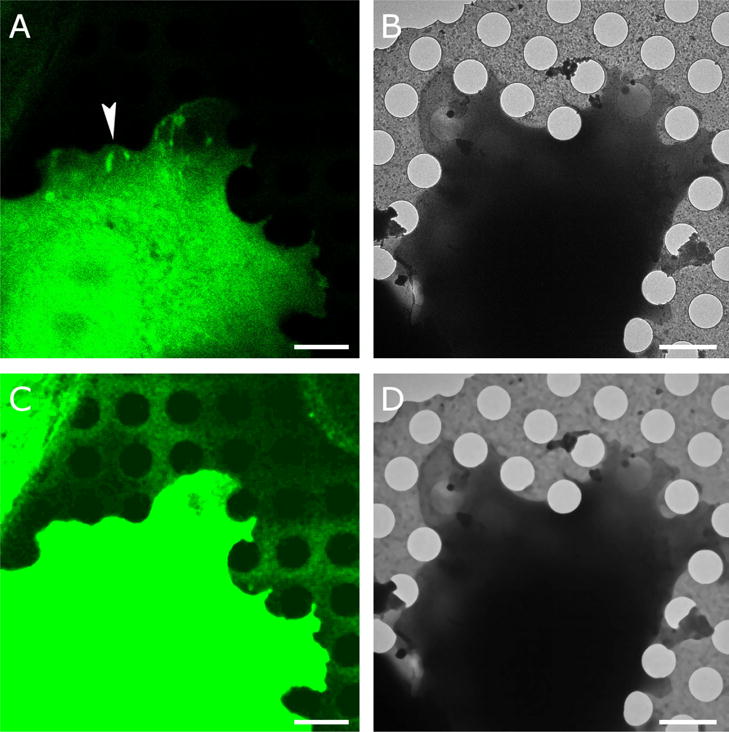
Figure 1. Correlative imaging.

A. An Olympus FV1000 confocal microscope equipped with a 60 × 1.35 UPlanSApo oil objective was used to capture Paxillin-GFP fluorescence (green) with a pixel size of 81 nm. Fluorescence was obtained at room temperature and the cell was plunge frozen after mild fixation (Anderson et al. 2016). B. Cryo-EM image of the same cell at 1500 × magnification resulting in 9.3-nm pixel size. The arrowhead in (A) points at a region with elongated spots of high paxillin fluorescence, indicative of focal adhesions, an actin-based assembly involved in cell adhesion to extracellular substrates. C. The holes in the carbon film are not visible in (A) but become visible after contrast adjustment and noise suppression with iterative median filtering. D. Iterative median filtering also suppresses background fluctuations in the cryo-EM image, allowing more accurate determination of the hole centers. Bars are 2 microns.
