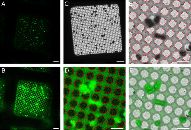
Figure 4. Correlating cryo-fluorescence and electron cryo-microscopy images.

A. Correlated R3045 ComGA:GFP fluorescence (green) of expressing Streptococcus pneumoniae recorded with a CorrSight cryo-microscope (FEI company). A 40 × objective was used with a camera pixel size of 161 nm. B. Enhanced cryo-fluorescence image showing the detectability of the holes in the carbon film. C. Cryo-EM image of the same field of view. The magnification is 500 × with 28-nm pixel size. D. Enlarged region of enhanced cryo-fluorescence with Hough transform circles (red) overlaid. E. Enlarged region of cryo-EM image with Hough circles (red) overlaid. F. Alignment of cryo-fluorescence (green) with cryo-EM image (grayscale) based on the centers of corresponding Hough circles. Bars are 5 microns.
