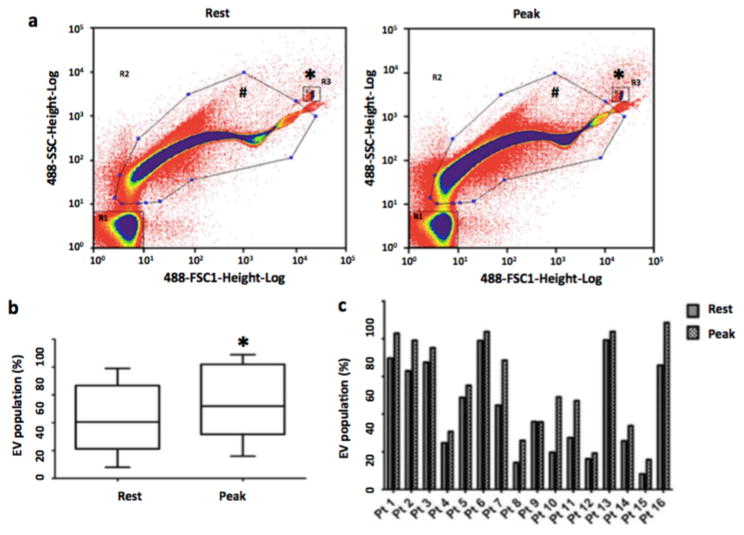
Figure 1. Acute exercise increases extracellular vesicle numbers in human subjects with cardiometabolic risk factors.
(a) Nano-FCM demonstrating EV profiles in patients in response to exercise during Exercise Stress Test. Representative nano-FCM profiles were shown at baseline (left) and at peak exercise (right). The rectangular gating (denoted by *) at the right upper quadrant denotes the fluorescent beads sized at 200 nm. EVs are in the gating denoted by hash symbol (b) Box-and-whisker plot demonstrating EV population (reported through the GeoMeans of the gated populations and expressed as percentage of the EV-gated population to all counted events) pooled from all the patient samples. (c) Bar graph demonstrating EV population (reported through the GeoMeans of the gated populations and expressed as percentage of the EV-gated population to all counted events) in individual patient (Pt) during resting state and at peak exercise. EVs, extracellular vesicles. *, P<0.05 (paired t-test, 2 tailed).

