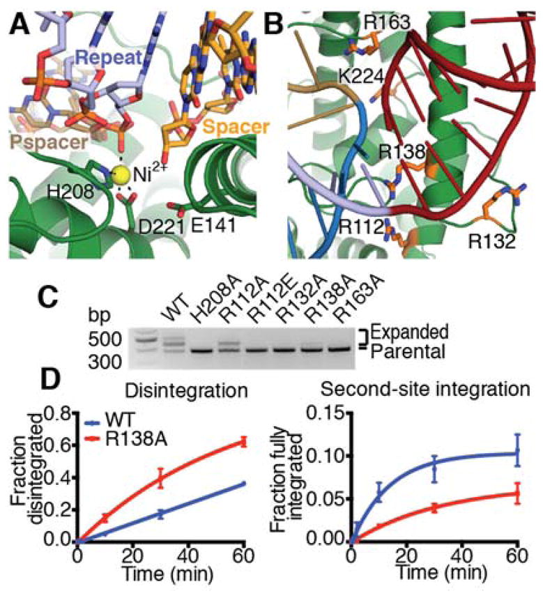
Fig. 4. Full-site integration requires a basic clamp around the active site.

(A) Metal coordination in the spacer-side active site. Active site residues, repeat, spacer, and protospacer (pspacer) are labeled, and coordination is shown as dotted lines. (B) View of basic residues surrounding leader-repeat junction. Basic residues in close proximity to the target DNA backbone on either side of the integration site are shown as sticks and colored orange. (C) Agarose gel of in vivo acquisition assay with indicated Cas1 mutants. H208A Cas1 is used as a negative control. (D) Quantification of disintegration and second-site integration time-course assays by wild-type and R138A Cas1. Mean and standard deviation of three independent experiments are plotted. Representative gels are shown in Figure S8.
