Fig. 4.
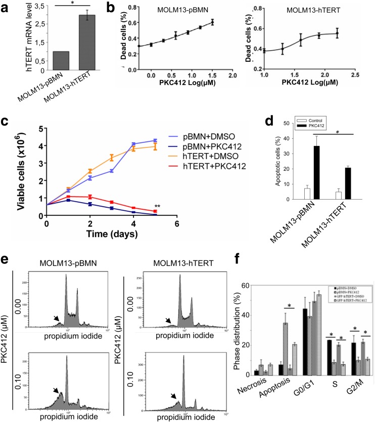
The attenuation of PKC412-induced apoptosis by ectopic hTERT expression in MOLM-13 cells. a MOLM-13 cells were infected with either control empty vector (pBMN) or hTERT-expressing vector to generate two sublines: MOLM-13-pBMN and MOLM-13-hTERT. The over-expression of hTERT in MOLM-13-hTERT cells was demonstrated using qPCR assay. b Cells were incubated with different concentrations of PKC412 for 48 h and IC50 then determined. Left panel: MOLM-13-pBMN cells and IC50 17.2 μM. Right panel: MOLM-13-hTERT cells, IC50 34.1 μM. c Cells were treated with 0.0125 and 0.1 μM PKC412 for 120 h, and viable cells were counted every 24 h. **P < 0.01. d, e hTERT-mediated attenuation of PKC412-induced apoptosis. MOLM-13-pBMN and MOLM-13-hTERT were treated with PKC412 at 0.1 μM and then analyzed for apoptotic cells using FACS. Representative FCCS graphs were shown in e, and the arrowheads point to the sub-G1 fractions (apoptotic cell population). *P < 0.05; ** P < 0.01. One-way ANOVA followed by LSD test was performed. f Cell cycle distributions in PKC412-treated cells. All the results shown were from three independent experiments

