Fig. 1.
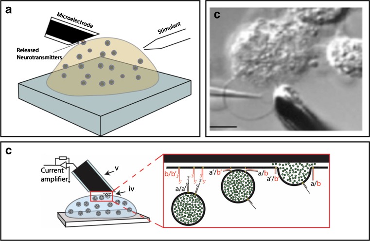
Here illustrates a few examples how the exocytosis process can be studied using live cells and synthetic cell models. a A schematic of an experimental set-up for amperometric measurement of exocytosis at live single cell [37]. b A DIC image of a cell model from bleb plasma membrane of a chromaffin cell, scale bar equals 10 μm [60]. c A schematic of a protein-free cell model using DNA-zipper molecules mimicking the SNARE protein complex role in catalyzing vesicle fusion and neurotransmitter release [81]

