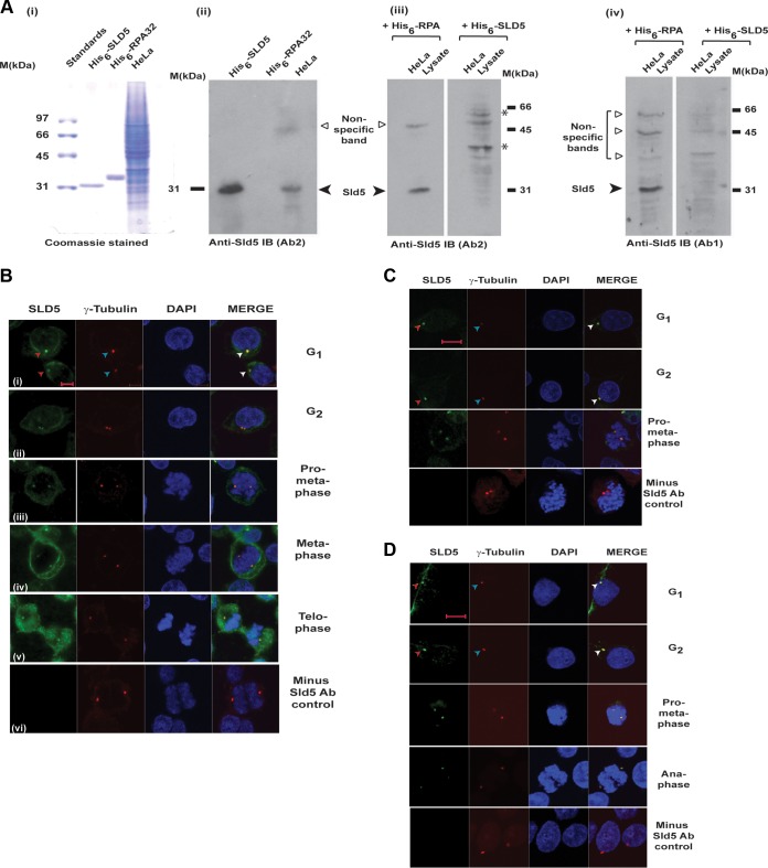
FIG 1.
Sld5 colocalizes with γ-tubulin at centrosomes. Sld5 localization to centrosomes was confirmed by immunofluorescence assays with multiple antibodies. (A) His6-tagged Sld5 protein expressed in E. coli was injected into rabbits to produce anti-Sld5 antibody. (i) His6-tagged Sld5 (0.5 μg) and His6-tagged RPA32 (0.5 μg) purified on a nickel-NTA column and 15 μg HeLa cell lysate were resolved by SDS-PAGE and stained with Coomassie blue. (ii) Alternatively, they were probed with Ab2 anti-Sld5 antibody. (iii) Ab2 anti-Sld5 antibody was incubated with 5 ng/μl His6-Sld5 or control His6-RPA protein, and the blots were developed with the same exposure time. Preincubation with His6-Sld5 but not His6-RPA protein led to the loss of Sld5 immunoblot (IB) signal observed at 31 kDa. Note that the nonspecific bands did not significantly change due to preincubation with His-Sld5 protein. Due to the presence of bacterial protein, some nonspecific sticking occurred (bands marked by asterisks), which was absent in the initial Ab2 immunoblot. (iv) Specificity of Ab1 antibody was demonstrated as explained for blot iii. (B) HeLa cells were prepermeabilized to remove the nuclear fraction of Sld5, followed by coimmunofluorescence assays with rabbit anti-Sld5 (Ab1) and mouse anti-γ-tubulin antibodies, in combination with anti-rabbit Alexa Fluor 488- and anti-mouse Alexa Fluor 555-conjugated antibodies, respectively. DNA was stained with DAPI. The right column is a merge of Alexa Fluor 488, Alexa Fluor 555, and DAPI images. (i to v) Cells in interphase (i and ii) and different phases of mitosis (iii to v). (vi) Immunofluorescence assays carried out in the absence of anti-Sld5 antibody, with other conditions and antibodies remaining similar, ruled out nonspecificity of secondary Alexa Fluor 488-conjugated antibody, as well as bleed-through of the Alexa Fluor 555 signal. (C and D) HeLa cells in different phases of the cell cycle were prepermeabilized to remove the nuclear fraction of Sld5, followed by coimmunofluorescence assay with either Ab3 anti-Sld5 antibody (C) or Ab2 anti-Sld5 antibody (D). Centrosomes are marked by arrowheads. Scale bars, 10 μm.

