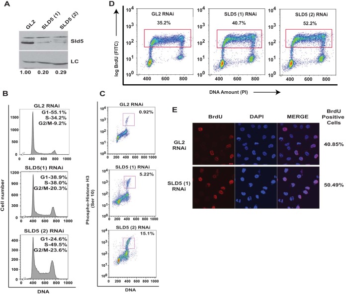
FIG 3.
Depletion of Sld5 leads to accumulation in M phase. (A) HeLa cells were transfected on three consecutive days with control GL2, SLD5 (1), or SLD5 (2) (targeting a different region of Sld5) siRNA, and the lysates were immunoblotted with anti-Sld5 antibody to confirm the specificity of RNAi-mediated depletion. LC, loading control showing equal protein loads in different lanes; the numbers indicate levels of Sld5 relative to control GL2 siRNA-transfected cells. (B and C) Transfected cells were harvested 24 h after the last transfection, followed by staining with propidium iodide alone (B) or in combination with anti-phospho-histone H3 (Ser 10) antibody (C), which marks the mitotic cells. The percentages in panel B show cell cycle distribution, while panel C shows cells in M phase. (D) Sld5 depletion causes S phase delay. Transfected cells were pulsed with BrdU for 30 min, followed by staining with anti-BrdU antibody conjugated to fluorescein isothiocyanate (FITC), along with propidium iodide (PI). The dot plots show BrdU incorporation (y axes) and DNA content (x axes), and the percentages of cells incorporating BrdU. (E) Transfected cells were pulsed with BrdU for 20 min, followed by staining with anti-BrdU (red) antibody. The coimmunofluorescence images display BrdU incorporation in different samples, while DNA was stained with DAPI (blue). Scale bar, 10 μm.

