FIG 7.
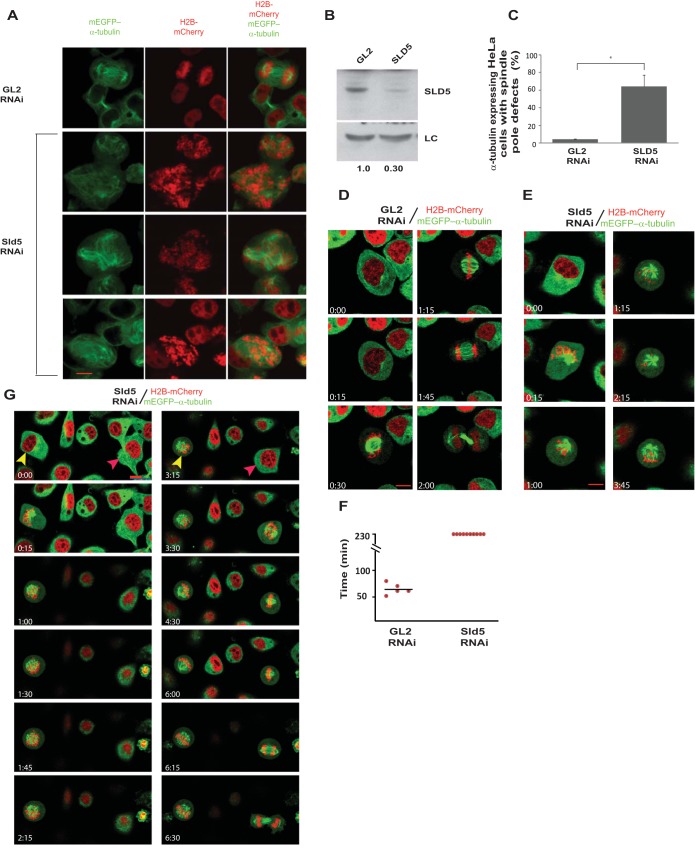
Sld5-deficient centrosomes fragment during chromosome congression. (A) HeLa cells stably coexpressing a red chromatin marker (H2B-mCherry) and a marker for microtubules (mEGFP–α-tubulin) were transfected on three consecutive days with control GL2 or SLD5 siRNA, followed by fixation for visualization of spindle pole defects. Multiple fields of SLD5 siRNA-transfected cells are shown. (B) Immunoblotting confirmed depletion of Sld5 in cells coexpressing H2B-mCherry and mEGFP–α-tubulin. LC, loading control showing equal protein loads in different lanes; the numbers indicate levels of Sld5 relative to control GL2 siRNA-transfected cells. (C) Quantification of spindle pole defects observed in GL2 or SLD5 siRNA-transfected cells coexpressing H2B-mCherry and mEGFP–α-tubulin. The data are represented as the means and SD of the results of two independent experiments, with more than 20 cells analyzed in each sample (*, P ≤ 0.05). (D and E) HeLa cells stably coexpressing H2B-mCherry and mEGFP–α-tubulin were transfected on three consecutive days with control GL2 or SLD5 siRNA, followed by live-cell imaging for almost 4 h. Selected frames at the indicated time points are shown (live-cell image capture is shown in Movies S1 and S2 in the supplemental material). Note that control cells progressed from interphase to cytokinesis, whereas SLD5 siRNA-transfected cells were arrested in an abnormal prometaphase until the end of the imaging period. (F) Quantification of the time taken by GL2 or SLD5 siRNA-transfected cells to progress from prometaphase to cytokinesis. Each point represents a single cell, while the mean is shown as a horizontal bar. Live-cell imaging is presented up to 230 min, at which time the Sld5-depleted cells were alive but had not progressed to anaphase. (G) HeLa cells stably coexpressing H2B-mCherry and mEGFP–α-tubulin were transfected on three consecutive days with SLD5 siRNA, followed by live-cell imaging. The captured images show a cell (yellow arrowheads) that did not complete mitosis and developed chromosomal and spindle pole defects. A small fraction of the cells (red arrowheads) completed mitosis, albeit slowly (mitosis started at time point 3:15 and was complete by 6:30). Note that almost all GL2 siRNA-transfected cells completed mitosis without developing chromosomal and spindle pole defects, establishing that the mitotic defects were due to Sld5 depletion. Scale bars, 10 μm.

