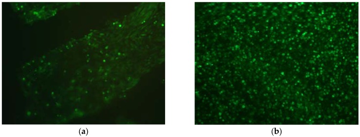
Figure 8.
Fluorescence live/dead staining of primary porcine chondrocytes growing on decellularized porcine cartilage five days after cell seeding. Overlay of green (live cells) and red (dead cells) channels; 200×. (a) Cells growing on non-crosslinked cartilage; (b) Cells growing on cartilage treated with 1% EGCG.

