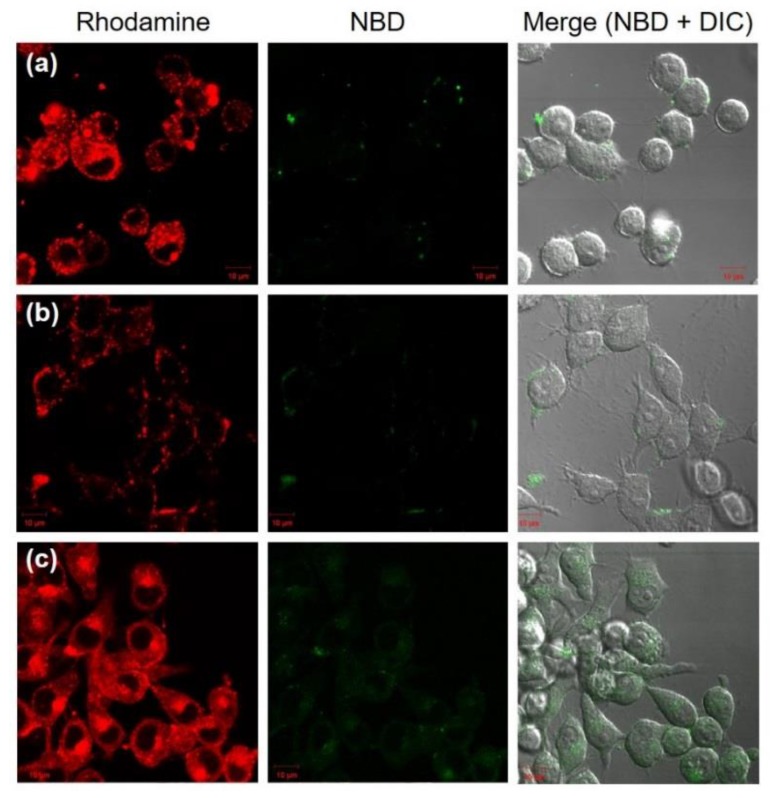
Figure 4.
Confocal laser scanning microscopic (CLSM) images of DC2.4 cells treated with DLPC liposomes (a), EYPC/deoxycholic acid micelles (b) and DLPC/deoxycholic acid micelles (c) for 5 h. Fluorescence of NBD-PE and Rh-PE upon excitation at 488 nm was observed using a CLSM. Scale bar represents 10 μm.

