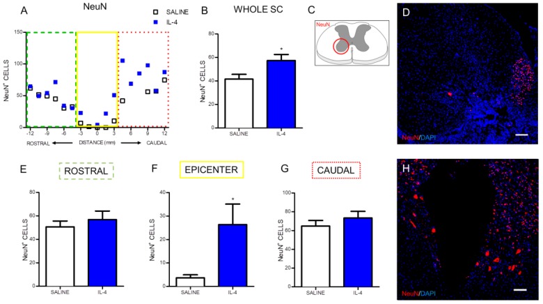
Figure 5.
IL-4 treatment increases the number of motor neurons in the ventral horns. Distribution of the NeuN+ cells along the rostrocaudal axis of the spinal cord (A); Quantification of NeuN+ cells in the whole spinal cord revealed a significant increase of motor neurons in IL-4-treated rats (B); A significant increase of motor neurons was also observed at the epicenter region of the spinal cord (F); while in the rostral (E) and at the caudal (G) areas no differences could be observed. Schematic image indicating areas where the analyses were performed (C); Representative images of positive staining for motor neurons of saline (D) and IL-4 treated (H) group. Values shown as mean ± SEM. * p < 0.05. Scale bar = 100 µm.

