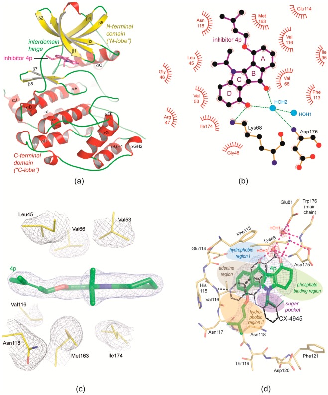
Figure 3.
The principle binding mode of 4p to protein kinase CK2. (a) Overview of the CK2α/4p complex structure. (b) 2D-projection of the non-covalent interactions between 4p and CK2α. (c) Hydrophobic packaging of 4p by non-polar side chains from the N-lobe (Leu45, Val53 and Val66), from the C-lobe (Met163 and Ile174) and from the interdomain hinge (Val116); the pieces of electron density were drawn with a cutoff level of 1 σ. (d) The ATP site of CK2α with bound 4p embedded in electron density (cutoff level 1 σ); for comparison the CK2 inhibitor CX-4945 was drawn with black C-atoms after superimposition of the protein matrices; the five regions of the ATP-site according to the protein kinase pharmacophore model of Traxler and Furet [63] are indicated by coloured patches. All parts of the figure were prepared with chain A of the low-salt CK2α1−335/4p structure (no. 2 of Table 1).

