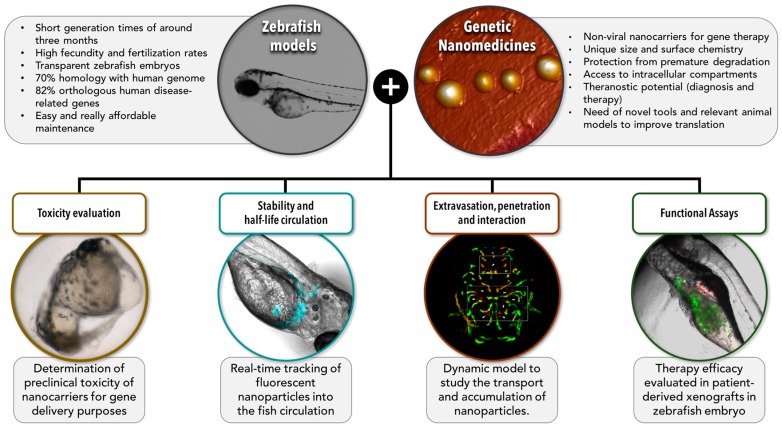
Figure 2.
Zebrafish as a model organism for preclinical studies of genetic nanomedicines. This scheme highlights the main characteristics of zebrafish as model organisms and the main advantages of nanomedicines for gene delivery. The scope of this review is summarized in the lower section of the figure where we have illustrated different ways in which zebrafish models can be extremely useful to help us understand the biological behaviour of genetic nanomedicines, and define better prototypes with improved opportunities of translation to a clinical setting. Zebrafish models would allow performing several assays of interest such as (i) evaluation of the toxicological profile, (ii) determination of the stability and half-life circulation of nanomedicines inyected in the fish circulation system, (iii) study of the ability of nanomedicines to extravasate, difuse, penetrate into the tumor, and interact with the targeted cells, and (iv) functional assays to test the potential and the efficacy of the proposed nanomedicines. The two images on top correspond to a zebrafish embryo (left), and to nanometric (~100 nm) lipidic nanoemulsions observed by atomic force microscopy (AFM) (right). Images in the low part of the figure correspond, from left to right, to 48 hpf malformed zebrafish embryo due to toxic effects of nanocapsules (image reproduced with permission from Teijeiro-Valiño et al. [88], fluorescent DiD-labelled lipidic nanoemulsions (blue) injected into the fish circulation system and observed under a fluorescence microscope (images adquired at 48 h post-injection), fluorescent nanoparticles (red) able to extravasate blood vessels (green) in a zebrafish model (image obtained by confocal microscopy by Zou et al. [133], and reproduced with permission), and fluorescent DiD-labelled lipidic nanoemulsions (red) able to interact with cancer cells (green) in xenotransplanted zebrafish embryos (HCT116-GFP) after yolk microinjection.

