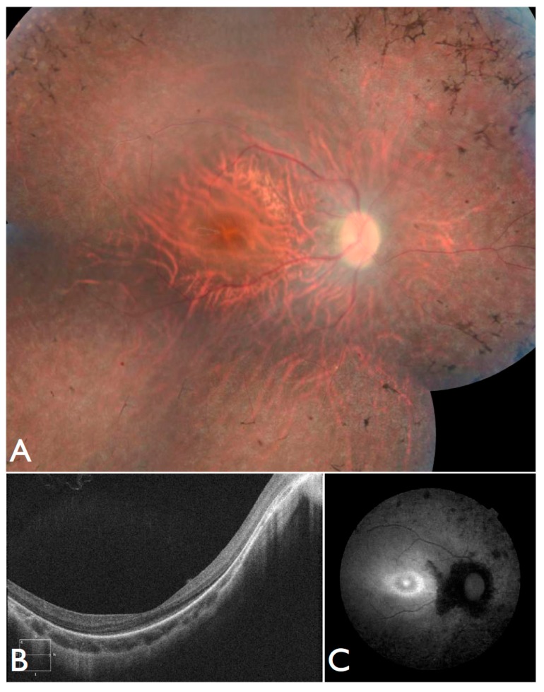
Figure 8.
Fundus and OCT images for FBP_53, solved by mutations in INPP5E. (A) Color fundus montages showing bilateral pale optic discs, attenuated retinal vessels, normal foveal reflexes, and mild desaturation of the fundus coloration with macular preservation and pigmentary disturbance at the periphery. (B) OCT scan and (C) Retinal autofluorescence.

