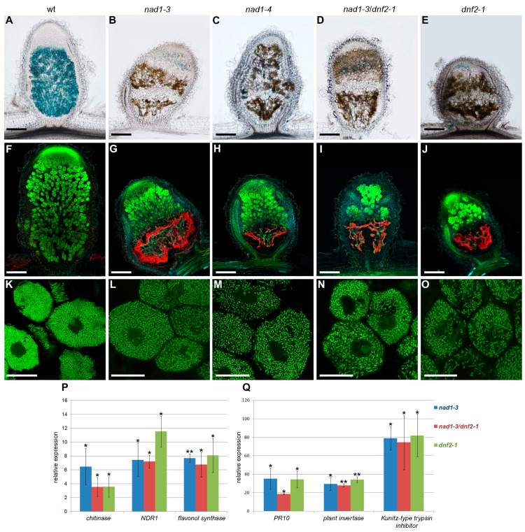
Figure 1.
Induction of defense responses in nodules with activated defense 1 (nad1), defective in nitrogen fixation 2 (dnf2) and nad1/dnf2 mutant nodules. The degree of rhizobial infection, bacteroid differentiation and the presence of brown pigmentation showing autofluoescence in symbiotically ineffective Medicago truncatula mutants supposed to be deficient in suppression of plant defense responses during the symbiotic interaction. Nodules were harvested 14 days post-inoculation (dpi) with Sinorhizobium medicae WSM419 expressing the lacZ marker gene. Nodule sections were stained for β-galactosidase activity (A–E) or stained with SYTO13 (F–O) and analyzed by light or confocal microscopy, respectively. Wild-type nodules showed the characteristic zonation of indeterminate nodules (A,F). Nodules of nad1-3 (B,G), nad1-4 (C,H), nad1-3/dnf2 (D,I), and dnf2 (E,J) displayed extensive brown pigmentation that corresponded to the area showing autofluorescence which is pseudocolored in red. Higher magnification revealed elongated bacteroids in the infected cells of the interzone of wild-type nodules (K). Elongated bacteroids were detected in the last layers of infected cells in mutant nodules (L,M,N,O), which indicates the initiation of bacteroid development in these nodules. Scale bars: (A–J) 200 μm, (K–O) 20 μm. (P,Q) Transcriptional activation of defense-related genes in the nodules of nad1-3, nad1-3/dnf2 and dnf2 mutants. The expression level of a chitinase, the NDR1, flavonol synthase, the PR10, a plant invertase and a Kunitz-type trypsin inhibitor gene was identified by quantitative reverse transcription PCR 14 dpi with S. medicae WSM419. The transcript levels were identified using three biological replicates and calculated relative to the expression detected in wild-type nodule. Error bars indicate ± standard error (SE). ** and *, p ≤ 0.01 and 0.05, respectively.

