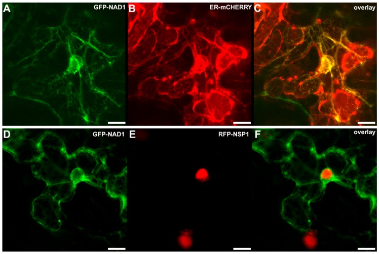
Figure 4.
Co-localization of NAD1 with localization control constructs in Nicotiana benthamiana leaf epidermal cells. The p35S::GFP-NAD1 and the ER marker p35S::ER-mCherry or the nucleus marker pUbq10::RFP-NSP1 constructs were co-transformed transiently into Nicotiana benthamiana leaves. The localization of the tagged proteins was imaged by confocal microscopy. The signal of GFP-NAD1 was detected in the endoplasmic reticulum (ER) network (A,D), which was confirmed by the co-localization with the mCherry signal targeted to the ER (B). The GFP-NAD1 is excluded from the nucleus but NSP1 shows nucleus localization (E). Panels C and F show overlay images of A and B, D and E, respectively. Scale bars: (A–I) 10 μm.

