Figure 4.
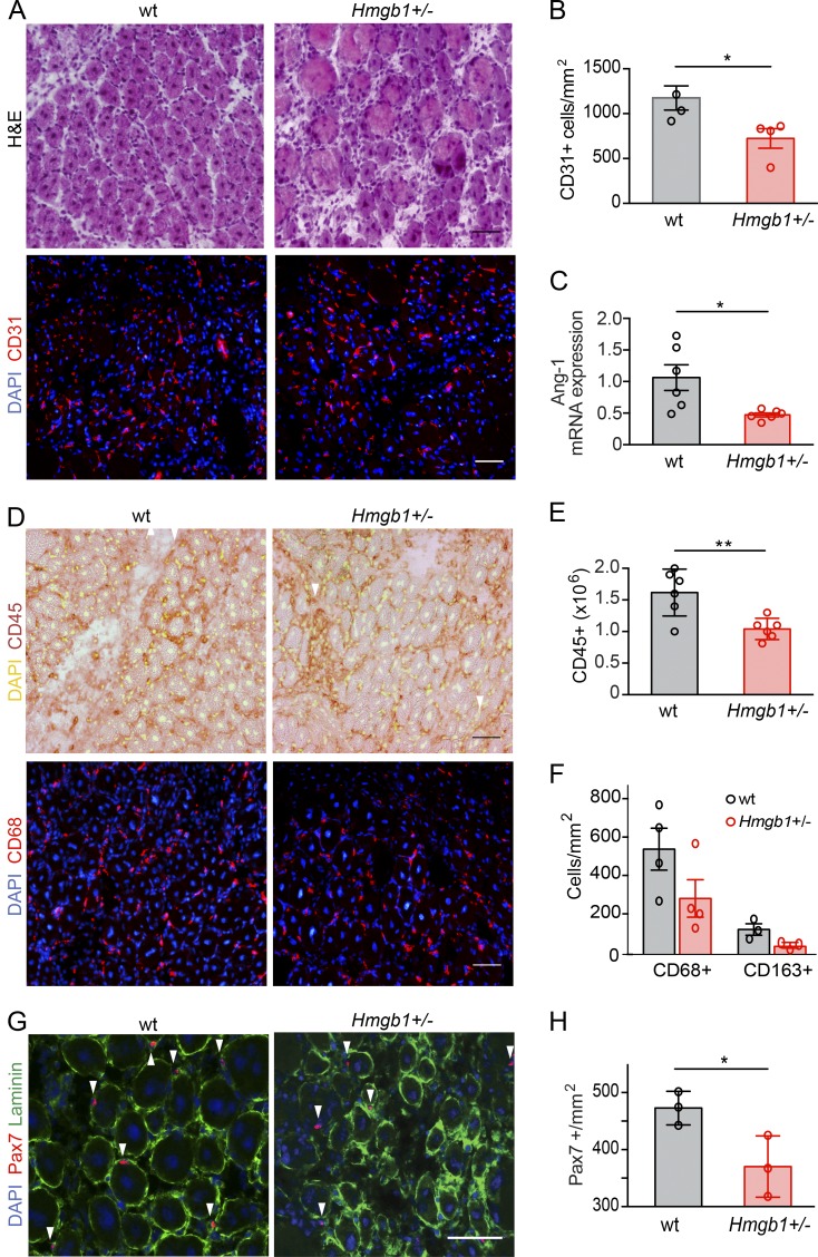
High expression of HMGB1 is required for optimal skeletal muscle regeneration. Muscle acute injury was induced by injection of Ctx in TA and/or triceps muscles of WT or Hmgb1+/− mice, and regeneration was assessed at day 5 after injury. (A) H&E and immunofluorescence staining (DAPI and CD31) of TA muscle sections from WT and Hmgb1+/− mice at day 5 after injury. Bars, 50 µm. (B) Quantification of CD31-positive cells/mm2 in TA muscles of WT and Hmgb1+/− mice at day 5 after injury. n = 4 mice per group of three independent experiments. (C) Quantitative PCR analysis of mRNA levels of angiopoietin-1 (Ang-1) in triceps at day 5 after injury. n = 6 mice per group of two independent experiments. (D) Representative immunostaining (DAPI and CD45, top; DAPI and CD68, bottom) of TA muscle sections from WT and Hmgb1+/− mice at day 5 after injury. Bars, 50 µm. (E) Quantification of CD45-positive cells isolated with immunobeads from injured muscles. n = 6 mice per group of two independent experiments. (F) Quantification of CD68-positive cells (n = 4 mice) and CD68/CD163-positive macrophages (n = 3 mice) in TA muscle sections from WT and Hmgb1+/− mice at day 5 after injury, two independent experiments. (G) Representative immunofluorescence staining for DAPI, laminin, and Pax7 in TA muscles from WT and Hmgb1+/− mice at day 5 after injury (Pax7-positive cells indicated with white arrowheads). Bars, 50 µm. (H) Quantification of Pax7-positive cells in TA muscles of WT and Hmgb1+/− mice at day 5 after injury. n = 3 mice per group of two independent experiments. In all panels, data are means ± SEM, and statistical significance was assessed with Student’s t test. *, P < 0.05; **, P < 0.01.

