Figure 6.
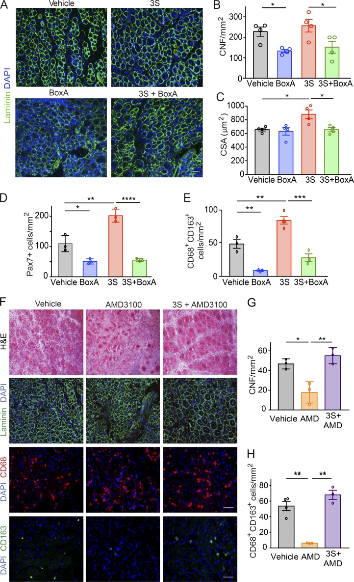
HMGB1 supports muscle regeneration via CXCR4. (A–E) Comparison of C57BL/6 mice treated with Ctx plus vehicle versus Ctx plus BoxA and Ctx plus 5 mg/kg 3S with or without 10 mg/kg BoxA. (A) Laminin and DAPI staining of TA muscle sections at day 5 after injury. Bar, 50 µm. Number (B) and CSA of centronucleated fibers (CNFs; C) in TA muscles at day 5 after injury. n = 4 mice per group, two independent experiments. (D) Quantification of Pax7+ cells in regenerating TA muscles at day 5 after injury. n = 3 mice per group, two independent experiments. (E) Quantification of CD68+CD163+ cells in sections of TA muscles at day 5 after injury. n = 4 mice per group, two independent experiments. (F–H) Comparison of muscles from mice receiving one single intramuscular injection of Ctx plus vehicle (PBS) versus Ctx plus 5 mg/kg AMD3100 with or without 5 mg/kg 3S. (F) Representative images of H&E and immunofluorescence staining for DAPI and laminin or CD68/CD163. Bars, 50 µm. (G) Number of CNFs in TA muscles at day 5 after injury. n = 3 mice per group, two independent experiments. (H) Quantification of CD68+CD163+ cells on sections of TA muscles at day 5 after injury. n = 3 mice per group, two independent experiments. Differences between groups were assessed with one-way ANOVA plus Tukey’s post-test. *, P < 0.05; **, P < 0.01; ***, P < 0.001; ****, P < 0.0001.

