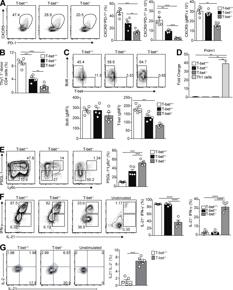
Figure 3.
T-bet is necessary for T cell development and their robust IFN-γ expression after LCMV challenge. T-bet+/+, T-bet+/−, or T-bet−/− Thy1.1+ Stg CD4+ T cells were transferred to Thy1.2+ B6 mice, followed by LCMV Armstrong infection 24 h later. Spleens were harvested 8 d p.i. (A) Representative flow cytometry plots of CXCR5 and PD-1 gating on PSGL-1loLy6Clo Tfh cells with bar graphs that summarize cell percentages, cell numbers, and CXCR5 MFI. (B) Percentage of donor cells that differentiated into Tfh cells in transfer recipients. (C) Representative intracellular Bcl6 and T-bet expression in PSGL-1loCXCR5hiPD-1hi Tfh cells with gMFI for both. (D) Quantitative RT-PCR for Prdm1 expression from sorted Tfh cells and control T-bet+/+ Th1 cells. (E) Representative flow cytometry plots of T-bet+/+, T-bet+/−, and T-bet−/− CD4+Thy1.1+PSGL-1loLy6Clo splenic Tfh cells from recipients, with a bar graph that summarizes percentages of cells. (F) Representative flow cytometry plots of intracellular IL-21 and IFN-γ staining of T-bet+/+, T-bet+/−, and T-bet−/− cells from recipient spleens, with bar graphs that summarize percentages of cells. (G) Representative flow cytometry plots of intracellular IL-21 and IL-2 staining of T-bet+/+ and T-bet−/− cells from recipient spleens, with a bar graph that summarizes percentages of cells. Data are representative of three experiments with three to five recipients per group. **, P < 0.01; ***, P < 0.001; ****, P < 0.0001 by Student’s t test. Error bars represent SEM.

