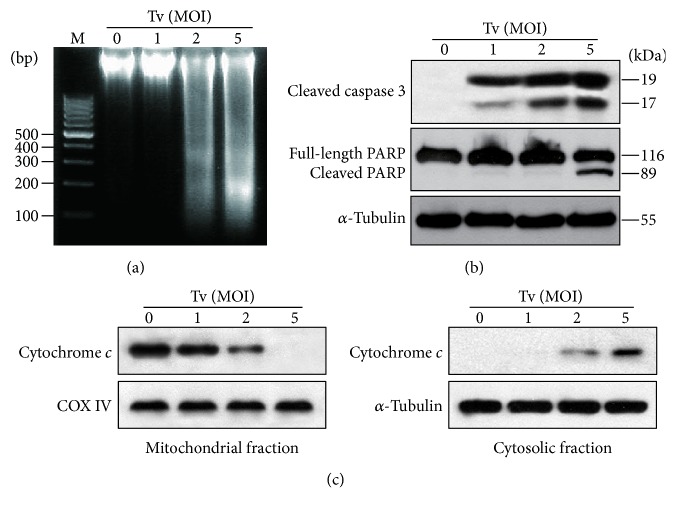
Figure 2.
Mitochondrial apoptosis was induced in SiHa cells following treatment with live T. vaginalis. (a) T. vaginalis induced DNA fragmentation, which is an indicator of apoptosis. (b) T. vaginalis induced the cleavage of caspase 3 and PARP. (c) T. vaginalis induced cytochrome c release from mitochondria into cytosol in parasite-burden-dependent manner. The quality of the fraction experiments was confirmed by assessing the presence of COX IV for the mitochondrial fraction and α-tubulin for cytosol fraction. The experiment was repeated three times with similar results. M: marker (100 bp DNA ladder); Tv: Trichomonas vaginalis.

