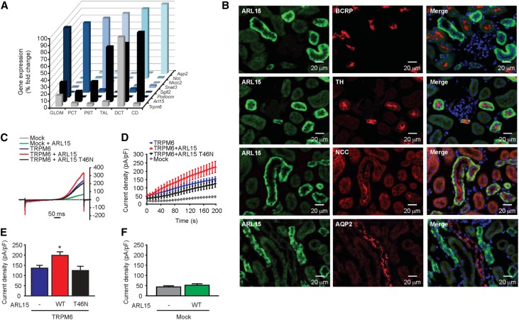
Figure 3.
ARL15 localizes in renal DCT regulating TRPM6 channel activity. (A) Gene expression analyses of Trpm6, Arl15, Podocin, Sglt2, Snat3, Nkcc2, Ncc, and Aqp2 in microdissected mouse nephron segments showed coexpression of Arl15 and Trpm6 in DCT. (B) Double immunofluorescence staining of mouse kidney cortex sections for ARL15 (in green) and BCRP (red); TH (red); NCC (red); or AQP2 (red) as markers of the proximal tubule, TAL, DCT, or collecting duct, respectively. For detection, the Alexa Fluor dye was used. (C) Typical current-voltage curves obtained from transfected HEK293 cells 200 seconds after break-in. Outwardly rectifying currents are observed in response to a 500 ms voltage ramp (from −100 to +100 mV) applied 200 seconds after break-in. (D) The average time development of the current density measured at +80 mV is shown (n≥10). The mock plus ARL15 condition is not shown for clarity reasons. (E) WT ARL15 (red, WT, n=47) but not the T46N ARL15 mutant (black, T46N, n=18) increased the whole-cell current density of TRPM6. Asterisk indicates significant difference with respect to the cells transfected with TRPM6 only (blue, “-,” n=44). One-way ANOVA followed by Tukey multiple comparisons post-test; P<0.05. (F) Transfection of HEK293 cells with WT ARL15 did not evoke a significant increase in whole-cell current density when compared with mock-transfected cells (n≥10, unpaired t test).

