Figure 1.
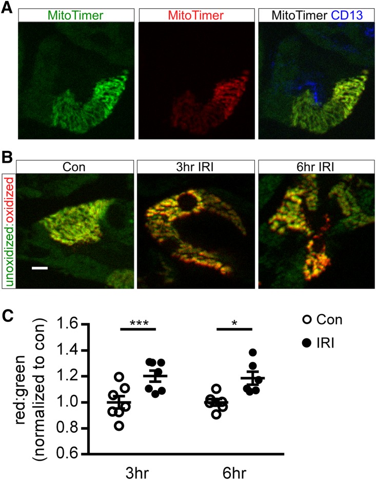
Outer medulla mitochondria become oxidized after IRI. Kidneys of PepCKCre MitoTimer mice were subjected to 26 minutes of unilateral ischemia and 3 or 6 hours of reperfusion. (A) Representative images of native MitoTimer fluorescence of individual green (left panel; unoxidized) and red (center panel; oxidized) channels and MitoTimer merged with CD13+ proximal tubule immunofluorescence (right panel) in control kidneys. (B) Representative images and (C) quantification of MitoTimer in the unoxidized and oxidized state in mitochondria in proximal tubule cells in the medulla. IRI indicates the kidney exposed to unilateral ischemia-reperfusion (operated kidney). Con, contralateral control (unoperated kidney). Scale bar, 10 μm. *P<0.05; ***P<0.001.

