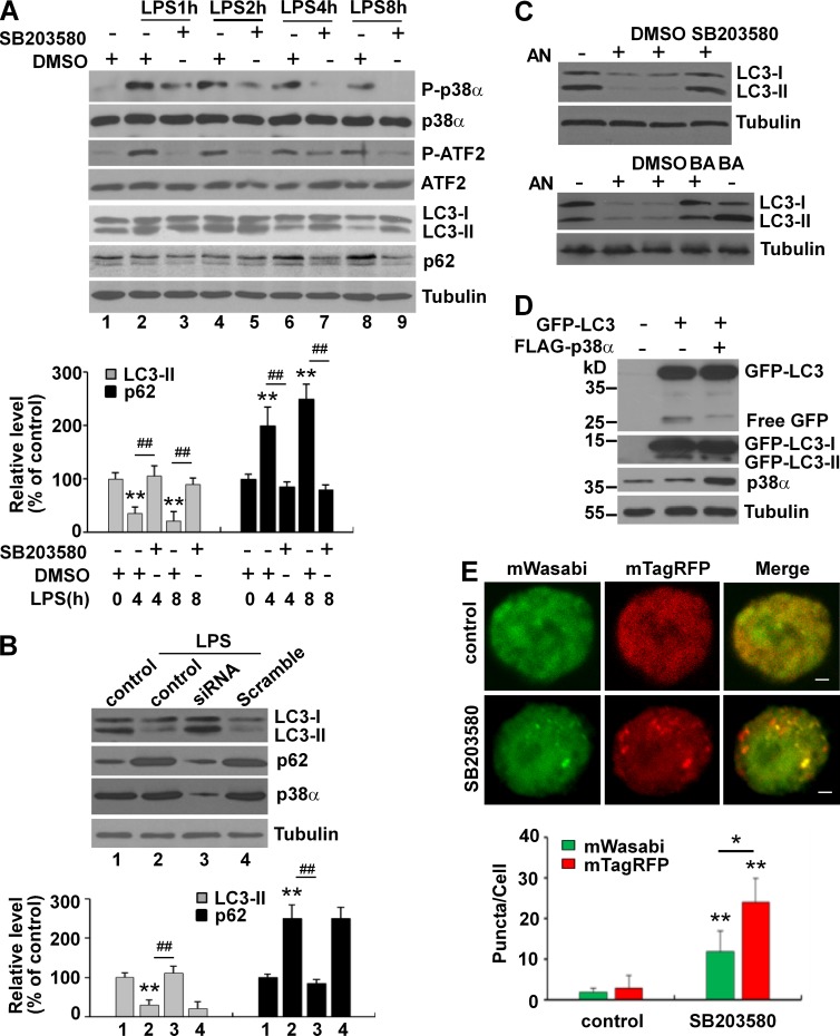
Figure 2.
LPS inhibits autophagy in BV2 cells through p38α MAPK. (A) p38 MAPK inhibitor SB203580 blocks LPS-induced inhibition of autophagy in BV2 cells. BV2 cells were treated with LPS (1 µg/ml) with or without SB203580 (10 µM) as indicated. The graph below shows relative changes of LC3-II and p62. (B) Knockdown of p38α MAPK blocks LPS-induced inhibition of autophagy in BV2 cells. BV2 cells were transfected with scramble or p38 MAPK–specific siRNA for 48 h and then treated with LPS (1 µg/ml) for 8 h. Whole-cell lysates were analyzed by immunoblotting. The graph below shows relative changes of LC3-II and p62. (C) The p38 MAPK activator AN inhibits autophagy in BV2 cells. BV2 cells were treated with AN (10 µM) with or without SB203580 (10 µM; top) for 8 h or were treated first with AN for 4 h and then with or without BA (100 nM; bottom) for another 4 h. (D) Expression of p38α MAPK reduces the conversion of GFP-LC3. HEK293 cells were transfected with GFP-LC3 or in combination with p38α MAPK for 24 h. Whole-cell lysates were analyzed by immunoblotting. (E) SB203580 enhances autophagy in BV2 cells under basal conditions. BV2 cells were transfected with tandem-fluorescently tagged LC3 (mTagRFP-mWasabi-LC3) for 24 h. Cells were treated with or without SB203580 (10 µM) for 8 h. *, P < 0.05; **, P < 0.01 versus control; ##, P < 0.01 versus LPS alone. Bars, 10 µm. n = 100. Experiments were repeated three times. Error bars show SD.

