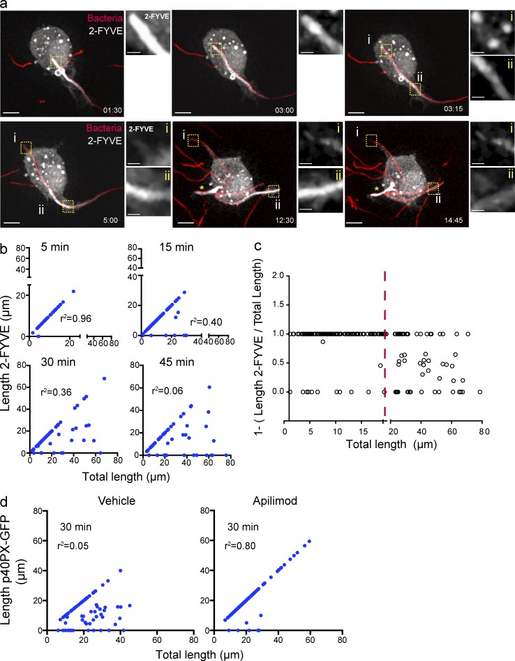
Figure 4.
PtdIns(3)P dynamics on the tPCs. (a) RAW macrophage expressing 2-FYVE-GFP (white), engulfing a filamentous bacteria (red), selected frames from Video 1. Images to the right of the main panels are magnifications of the framed regions in the main panels depicting 2-FYVE-GFP in tPCs. For the first two images, the area magnified corresponds to the most distal tip of the tPC containing filamentous bacteria. In the 4 last micrographs (times 3:15 to 14:45), panel i is the most distal tip of the tPC and ii is the proximal tPC. Bars: 5 µm; (enlarged areas) 1 µm. (b) RAW macrophages expressing 2-FYVE were challenged with filamentous bacteria and fixed after 5, 15, 30, and 45 min of phagocytosis. The length of filamentous bacteria positive for 2-FYVE was plotted against the bacterial length internalized at each time point. The corresponding correlation coefficients (r2) were calculated. (c) Regions of tPCs that surpass a length of 20 µm (red line) tend to be devoid of 2FYVE-GFP. The ratio of 2FYVE-GFP–positive length to the total length of the tPC is shown. A ratio of 1 indicates that the entire tPC is decorated with 2FYVE-GFP, a ratio <1 indicates that a portion is devoid of this probe, and a ratio of 0 indicates that the tPC is completely divested of the probe. Data from three independent experiments (n = 30 for each time point). (d) RAW macrophages expressing p40PX-GFP, treated with 10 nM apilimod or 0.1% DMSO (vehicle) for 1 h, were allowed to engage in phagocytosis of pHrodo-conjugated filamentous bacteria for 30 min of phagocytosis. The length of filamentous bacteria positive for p40PX-GFP was measured in three independent experiments, n = 30 for each condition, and plotted as described in panel b.

