Figure 9.
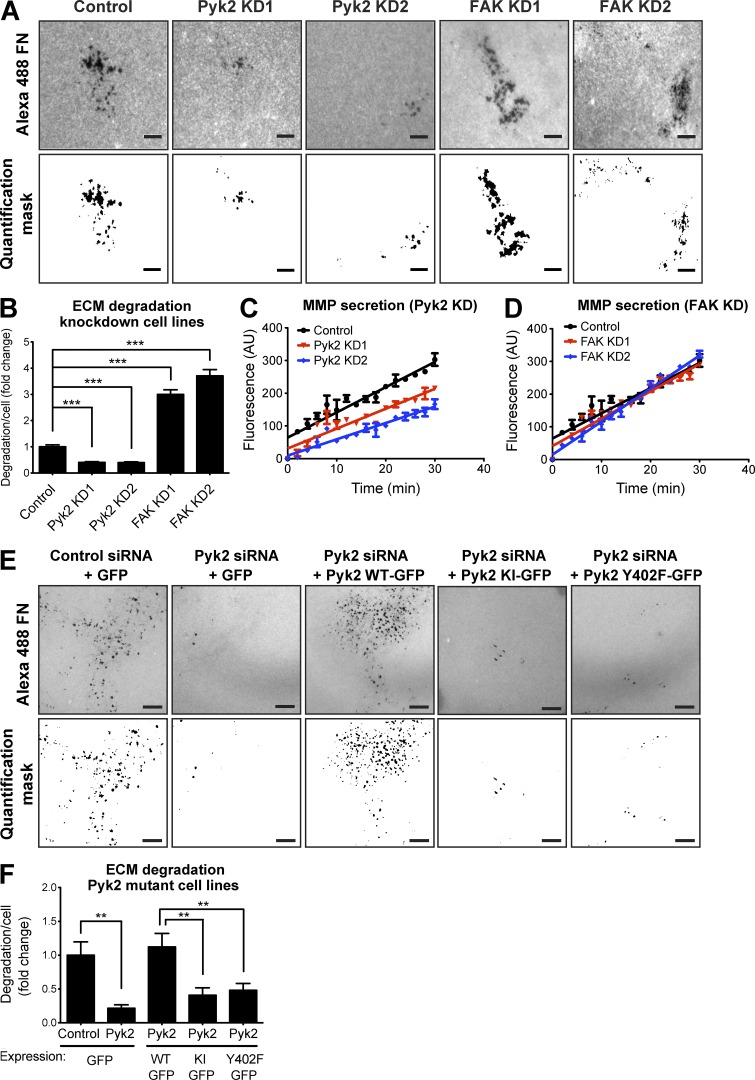
Pyk2 and FAK oppositely regulate ECM degradation by breast cancer cells. (A) MDA–MB-231 cells stably expressing control, Pyk2 shRNA, or FAK shRNA were plated on Alexa Fluor 488 FN/gelatin matrix and allowed to degrade for 24 h. Shown are representative images (left) and quantification masks (right) of degradation areas. (B) Quantification of matrix degradation by control and knockdown (KD) cells. n = 280–475 fields per group from three independent experiments. (C and D) MDA–MB-231 cells expressing control, Pyk2 shRNA, or FAK shRNA were incubated in serum-free medium in the presence of a fluorogenic substrate peptide, and their MMP activity was measured over 30 min. Shown are linear regression graphs. n = 8–10 samples per group from three independent experiments. (E and F) Representative images (E) and quantification (F) of matrix degradation by cells expressing WT Pyk2-GFP, kinase-inactive (KI) Pyk2-GFP, or Y402F-Pyk2 GFP and treated with control or Pyk2 siRNA. Bars, 10 µm. n = 42–73 cells per group from three independent experiments. **, P < 0.01; ***, P < 0.001. Error bars represent SEM.

