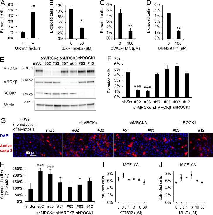
Figure 2.
Apoptotic epithelial extrusion relies on myosin contraction and MRCKα. (A) Epithelial extrusion, measured by counting the number of cellular bodies released in the culture medium from confluent MCF10A epithelium, is significantly increased by removal of growth factors for 16 h. (B) Treatment with the tBid inhibitor (50 µM) caused a decrease in the number of extruded cells after growth factor deprivation. (C) The same effect was recapitulated by treatment with the caspase inhibitor zVAD-FMK (100 µM). (D) Inhibition of myosin contraction by blebbistatin (100 µM) hampered epithelial extrusion. (E) Efficiency of MRCKα, MRCKβ, and ROCK1 silencing shown by immunoblot. (F) The quantification of extrusion in cells stably silenced for MRCKα, MRCKβ, or ROCK1 shows that only MRCKα is able to significantly impair epithelial extrusion from confluent cells compared with control shRNA (shScr). In these experiments, error bars represent the SEM. (G) Evaluation of apoptotic cells present in confluent epithelial layer 16 h after apoptotic induction detected by anti–cleaved caspase 3 antibody (in red). Signal detected in the epithelial layer is the sum of not-yet-extruded cells and apoptotic bodies resulting from faulty extrusion. (H) Quantification of apoptotic bodies (size 1.6–12.5 µm2) shows that extrusion-defective MRCKα silenced cells are forced to conclude apoptosis within the epithelial layer. (I and J) Extrusion assay in presence of different concentrations of Y27632 (I) and ML-7 (J) shows that apoptotic epithelial extrusion in MCF10A is independent of ROCK and MLCK. Error bars represent SEM (n = 3). *, P < 0.05; **, P < 0.01; ***, P < 0.001.

