Figure 6.
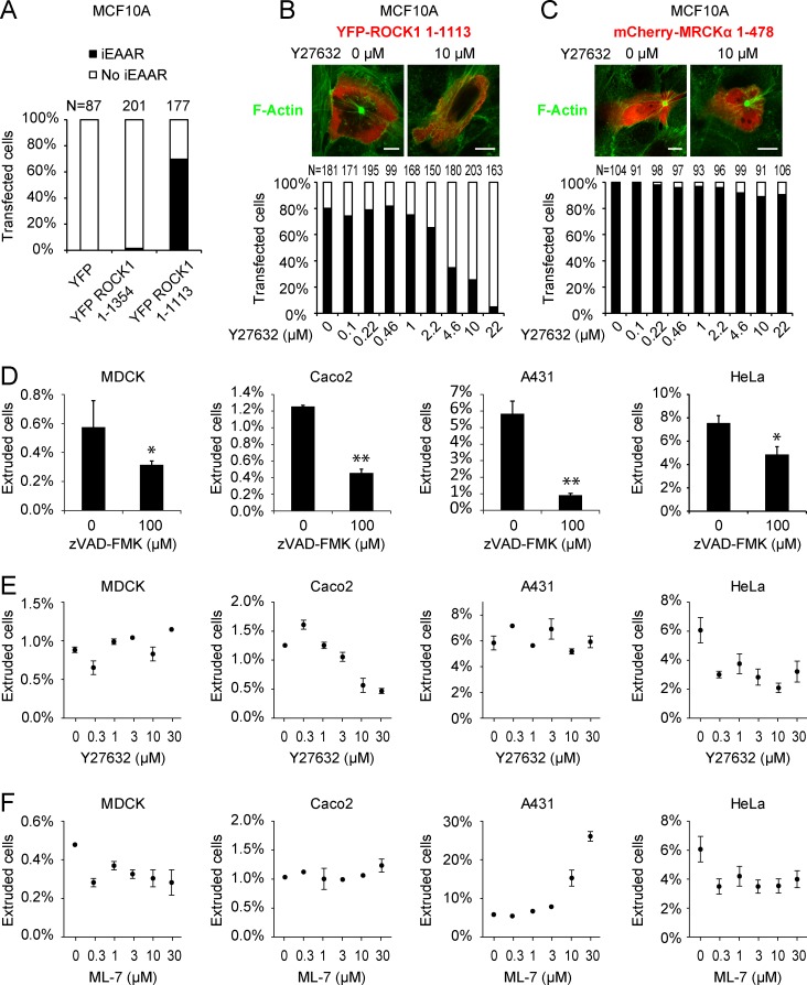
ROCK1 role in iEAAR assembly and in the regulation of apoptotic epithelial extrusion. (A) Percentage of MCF10A cells that show iEAAR formation after transfection with full-length YFP–tagged ROCK1 (YFP-ROCK1 1–1,354), with its apoptotic cleavage fragment (YFP-ROCK1 1–1,113) or YFP alone. Only YFP-ROCK1 1–1,113 is able to induce iEAAR assembly. (B) Treatment with Y27632 prevents iEAAR formation in MCF10A cells transduced with YFP-ROCK1 1–1,113. (C) Treatment with Y27632 fails to prevent iEAAR formation in MCF10A cells transfected with mCherry-MRCKα 1–478. Bars, 10 µm. (D) Apoptotic epithelial extrusion from confluent layer of HeLa, Caco2, MDCK, and A431 cells is prevented by the caspase inhibitor zVAD-FMK (n = 3). (E) The role of ROCK kinase activity in epithelial extrusion is shown in different cell lines by treating cells with different concentrations of Y27632 during the extrusion assay (n = 3). (F) The role of MLCK kinase activity in epithelial extrusion is shown the different cell lines by treating them with different concentrations of ML-7 during the extrusion assay (n = 3). *, P < 0.05; **, P < 0.01. Error bars represent SEM (n = 3).

