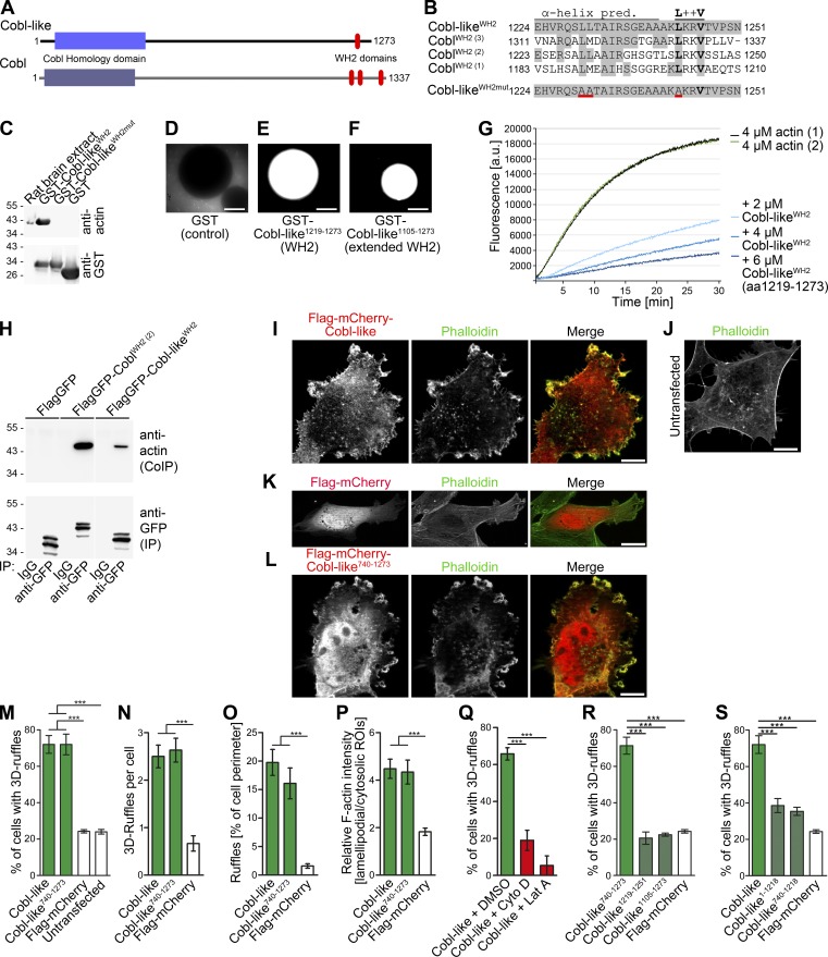
Figure 1.
Cobl-like is a WH2 domain–containing, G-actin–binding protein promoting F-actin–driven shape changes of COS-7 cells. (A) Scheme of murine Cobl-like in comparison to the actin nucleator Cobl. (B) Alignment of the predicted Cobl-like WH2 domain and a mutated (red underlining) version thereof with the three WH2 domains of Cobl. (C) Immunoblotting analyses of precipitations of endogenous actin from rat brain extracts (input) with immobilized Cobl-like WH2 domain, a mutated version of this domain (L1230A, L1231A, and L1243A; WH2mut), and GST as control. (D–F) Beads with immobilized GST-Cobl-like1219–1273 and GST-Cobl-like1105-1273 incubated with rat brain extracts supplemented with fluorescent actin and an energy-regenerating system showing merely a recruitment of fluorescent G-actin (but no formation of F-actin structures at the bead surfaces). Bars, 50 µm. (G) Pyrene-actin assays showing a dose-dependent suppression of spontaneous F-actin formation by Cobl-like1219–1273. (H) Specific coimmunoprecipitations of endogenous actin with Cobl WH2 (2) and the Cobl-like WH2 domain. White line, lanes omitted. (I–L) Maximum intensity projections (MIPs) of ApoTome images of (Flag-tagged) mCherry-Cobl-like (I) and mCherry-Cobl-like740–1273 (L) and F-actin in F-actin–rich membrane-ruffles in COS-7 cells. Untransfected cells (J) and Flag-mCherry–expressing cells (K) are shown as controls. Bars, 10 µm. (M–P) Quantitative evaluations of the Cobl-like and Cobl-like740–1273–induced phenotype of 3D ruffle formation by scoring the percentage of cells with such structures (M) and additional analyses substantiating the phenotype by further quantitative evaluations, such as the number of 3D ruffles per cell (N), ruffles in percentage of cell perimeter (O), and the ratio of lamellipodial versus cytosolic F-actin (P). (Q) Scoring of cells with the Cobl-like–induced 3D ruffles phenotype on latrunculin A (Lat A) and cytochalasin D (Cyto D) incubation, respectively. DMSO, solvent control. (R and S) Quantitative analyses of the molecular requirements of Cobl-like–induced 3D ruffle formation showing that the WH2 domain is not sufficient (R) but critical (S). Data are mean ± SEM, one-way ANOVA plus Tukey’s post test. ***, P < 0.001.

