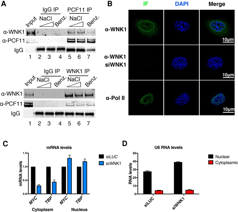
Figure 1.

WNK1 is present in the nucleus and interacts with PCF11. (A) Co-IP experiments of PCF11 and WNK1. Immunoprecipitation of PCF11 (top panel) and WNK1 (bottom panel). Lane 1 corresponds to 5% input. Lanes 2–4 show mock immunoprecipitation using rabbit IgG (negative control). Lanes 2 and 5 show immunoprecipitation with 150 mM NaCl, and lanes 3 and 6 show immunoprecipitation with 250 mM NaCl. Lane 7 shows immunoprecipitation with 150 Mm NaCl after incubation with benzonase to test for nucleic acid dependence. (B) Superresolution microscopy and detection of WNK1 kinase. Pol II staining was used as a control for nuclear localization. The same pattern was observed in all imaged cells. n = 20. Data are from two biological repeats. The same laser intensity was used for all images. (C) Cytoplasmic and nuclear levels of MYC and TBP mRNA in control and WNK1-depleted cells. Data are from biological repeats. Error bars represent standard error of mean (SEM). Data were normalized to siRNA targeting luciferase (siLUC). (D) U6 RNA levels measured with RT-qPCR in nuclear and cytoplasmic extracts.
