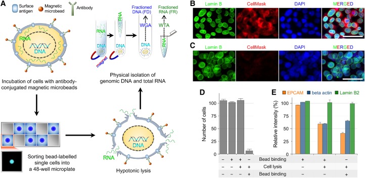
Figure 1.
Principles of the SIDR method. (A) Schematic of the SIDR method. (WGA) whole-genome amplification; (WTA) whole transcriptome amplification. (B,C) Immunostaining of the nucleus after cell lysis. Fluorescence images of MCF7 cells in isotonic (B) and hypotonic (C) conditions. The nuclear lamina, plasma membrane, and nucleus were stained by the Alexa 488-labeled anti-Lamin B2 antibody (green), CellMask (red), and DAPI (blue), respectively. (D) The effect of cell lysis on the recovery rate of cells. The recovery rate dramatically depended on whether anti-EPCAM antibody-conjugated microbeads were bound to cells before or after cell lysis as indicated at the bottom of the graph. Approximately 100 MCF7 cells underwent bead binding and/or cell lysis and were magnetically recovered, except for control cells that were not bound to microbeads. The plot shows the number of cells recovered (n = 3). (E) The effect of bead binding on the solubilization of the EPCAM protein. The levels of EPCAM, beta actin, and Lamin B2 proteins in cell lysates not solubilized during cell lysis were measured by Western blot (n = 3).

