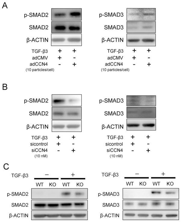
Fig. 3.
The effect of CCN4 on TGF-β3-SMAD signaling pathway in vitro. hBMSCs were transduced with adCCN4 (A) or siCCN4 (B) for 2 days. (A, B) Transduced-hBMSCs in monolayer culture were treated with 10 ng/mL of TGF-β3 for 10 min, and cellular proteins were collected for detection of SMAD2/3 activity. β-ACTIN was used as protein loading control. (C) Chondrocytes were collected from mandibular condylar cartilage of newborn WT and Ccn4-KO mice, and stimulated with TGF-β3 for 10 min. The cellular proteins were collected for analysis of TGF-β3-induced phosphorylation of SMAD2/3. SMAD2/3 activity was decreased in chondrocytes derived from Ccn4-KO mice, compared to WT mice.

