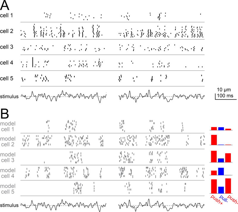
Figure 8. Different aPN3 neurons encode diverse temporal features of a wind-like stimulus.
A) Spiking in six aPN3 neurons evoked by a ”wind”-like stimulus. The stimulus (bottom) was constructed so that its power spectrum emulates that of antennal movements evoked by wind (dotted line is zero displacement, i.e. the resting position of the antenna). Two snippets of this stimulus are shown.
B) Responses of six model aPN3 neurons to the same stimulus. This is a subset of the model cells in Figure 6, and their parameters were fit only to the data in Figure 6; they are not intended to match the individual cells in (A). Nonetheless, each of these cells is selective for different temporal features of the “wind”-like stimulus, and in this regard they resemble the cohort of cells in (A), which are also each selective for different temporal features. Here we explicitly model spikes, rather than simply modeling the aPN3 neuron membrane potential. The transformation from voltage to spikes in the model is given by a differentiating function, in order to create the characteristic burstiness of aPN3 neurons (see Supplemental Experimental Procedures).

