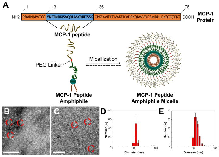
Figure 1.
Preparation and characterization of MCP-1 PAMs. (A) Schematic representation of MCP-1 PAMs. Synthetic MCP-1 peptides corresponding to the CCR2-binding motif (residues 13–35) of the MCP-1 protein were conjugated with DSPE-PEG(2000) to form MCP-1 PAs. (B) Representative TEM images of MCP-1 PAMs and (C) scrambled PAMs. Scale bar = 100 nm. (D) Particle size distribution of MCP-1 PAMs and (E) scrambled PAMs measured by DLS.

