
Figure 1. Specialized Membrane-Based Tubular Structures Enable Cell–Cell and Mitochondria–Mitochondria Information Transfer.
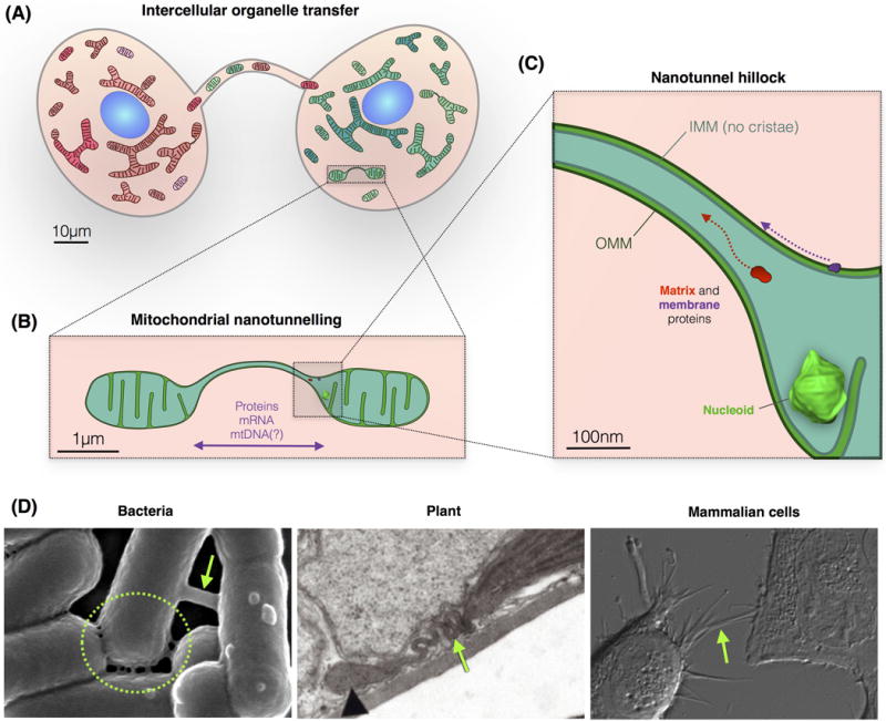
(A) Mammalian cell–cell exchange of organelles, vesicles, and soluble molecules occurs through cytonemes, nanotubes, and microtubules. (B) Within cells, mitochondria form similar tubular structures with contiguous outer and inner mitochondria membranes, and a continuous matrix space allowing the selective diffusion of specific molecular components. (C) Schematic of the nanotunnel junction, or ‘hillock’, showing the continuity of mitochondrial compartments. Nucleoid drawn to scale, see also Figure 2. (D) (Left) Scanning EM of intercellular nanotubes connecting PY79 bacteria [19]. (Center) Transmission EM of a tubular stromule extending from a chloroplast in a mesophyll cell of Arabidopsis thaliana [53]. (Right) Differential interference contrast (DIC) imaging of human HEK293 cells with cell–cell membrane protrusions. Abbreviations: EM, electron microscopy; IMM, inner mitochondrial membrane; mtDNA, mitochondrial DNA; OMM, outer mitochondrial membrane.