Figure 1.

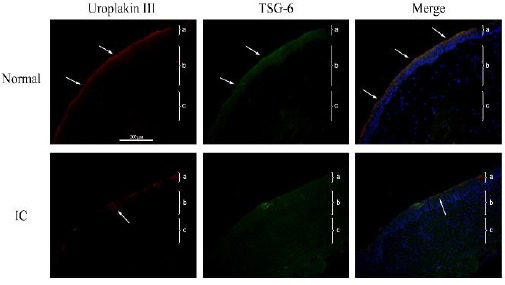
Representative immunofluorescence results (red, uroplakin III; green: tumor necrosis factor-inducible gene 6 (TSG-6); blue: DAPI) of the bladders of control human subjects (top row) or patients with bladder pain syndrome/interstitial cystitis (BPS/IC) (bottom row) (200×). Each tissue layer is labeled with a bracket “}” to indicate its range. a: transitional cell epithelium, b: lamina propria mucosae, c: muscle layer
Top row: note continuous expression of uroplakin III on the surface of the bladder epithelium (arrow), TSG-6 expression in and on the surface of the bladder epithelium (arrow), and significant overlapping expression of these 2 proteins in the merged image. Bottom row: note discontinuous expression of uroplakin III on the surface of the bladder epithelium with a focal deficit (arrow), and higher TSG-6 expression in the bladder epithelium, lamina propria mucosa, and tunica muscularis layers
