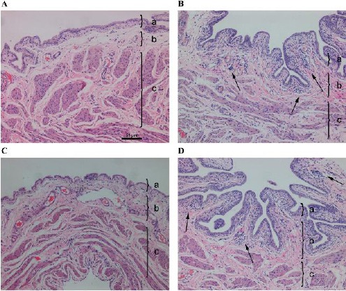
Figure 2.

Representative hematoxylin & eosin staining results of the bladders of rats in the control 1W group (A), hyaluronidase (HAase) 1W group (B), HAase 1W/ tumor necrosis factor-inducible gene 6 (TSG-6) group (C), and HAase 1W/PBS group (D) (100×). Each tissue layer is labeled with a bracket “}” to indicate its range. a: transitional cell epithelium, b: lamina propria mucosae, c: muscle layer. Note the focal deficit of bladder epithelium and infiltration of inflammatory cells in the submucosa and muscularis (arrow in B) and infiltration of inflammatory cells (arrow in D)
