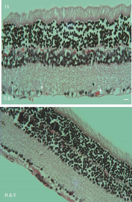
Figure 1.

Photomicrographs of the retina in diabetic group (1A) revealed histopathological changes such as ganglion cell layer shrinkage (arrow) and new vessel formation (arrowhead). Treatment with JRL and metformin ameliorated the dramatic histological alternations (1B) (stained with hematoxylin and eosin; original magnification: ×400, bar: 100µm)
