Figure 2.
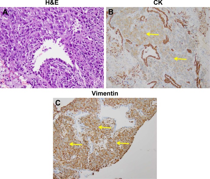
Photomicrographs showing the representative histologic appearance of pulmonary blastoma.
Notes: (A) Malignant epithelial elements in undifferentiated mesenchymal stroma stained by H&E (200×). (B) The malignant glandular component was diffusely positive for the epithelial marker CK (40×). (C) The stromal blastematous malignant component was diffusely positive for mesenchymal stromal marker vimentin (100×). The yellow arrows indicate the positive cells stained by relevant markers.
Abbreviations: CK, cytokeratin; H&E, hematoxylin–eosin.

