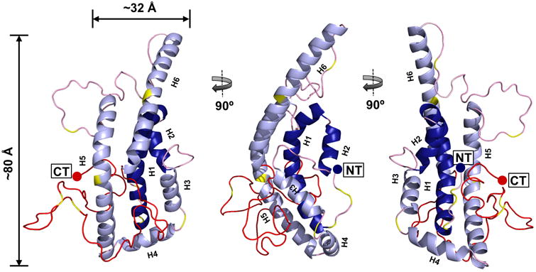
Figure 3. All atom model of full-length, lipid-free monomeric apoA-I.

The final time-averaged model is shown where dark blue represents N-terminal helical α-helical domains H1 and H2, light blue represents α-helical domains H3, H4, H5 and H6, pink represents random coil, red represents the C-terminal 59 residues absent in the crystal structure and yellow shows the position of prolines on each model.
