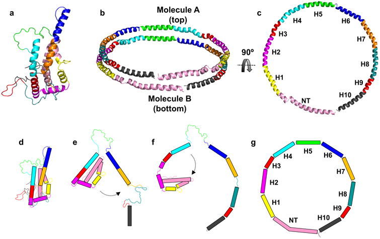
Figure 7. Hypothesis on lipid-binding and relationship to apoA-I arrangement on discoidal HDL.

Panel (a): The consensus model of full-length apoA-I. Panel (b): Proposed arrangement of two molecules of apoA-I on discoidal HDL; the “double-belt” model. Panel (c): Molecule B from the double-belt model rotated 90° (top view). Panel (d): Cartoon representation of the time-averaged model in its folded state. Panel (e): Dissociation of helix 6 and C-terminus from the lipid-free model and formation of a helical region upon interaction of the CT with lipid. Panel (f): formation of additional alpha-helical segments upon lipid filling and unfurling of the N-terminal bundle. Panel (g): Endpoint as a belt on an HDL particle. The model and cartoon is colored based on alpha-helical repeats identified and reported by Segrest et. al. 49. Rectangles in panels (d-g) represent alpha-helical segments.
