Figure 2.

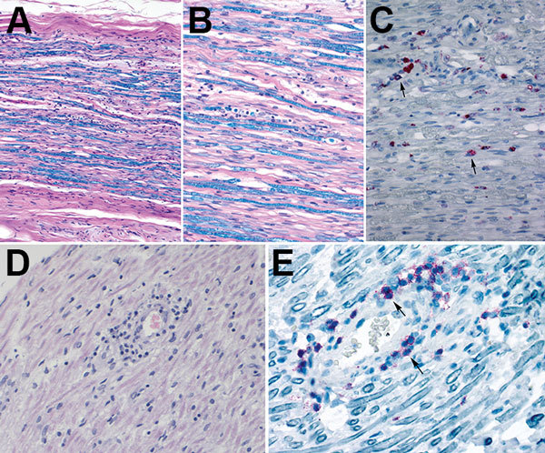
Histopathologic evaluation of tissue specimens collected postmortem from a patient with Guillain-Barré syndrome (acute demyelinating inflammatory polyneuropathy variant) and Zika virus infection, Puerto Rico, 2016. A, B) Luxol fast blue-periodic acid-Schiff myelin stain of sciatic nerves show patchy myelin loss and variable inflammation. Original magnification x10(A) and x20(B). C) Detection of CD68–positive cells (microphages) by immunohistochemistry (arrows) in sciatic nerve. Original magnification x20. D) Hematoxylin and eosin stain of cranial nerve IV shows perivascular lymphocytic infiltrate. Original magnification x20. E) Detection of T-lymphocytes by immunohistochemistry (arrows) in the same area where lymphocytic infiltrates were observed by hematoxylin and eosin stain. Original magnification x40.
