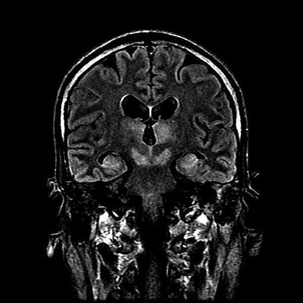
Figure 2.

Magnetic resonance imaging of brain of index patient 66 days after double lung transplantation, Hong Kong, China. Coronal FLAIR (FLuid Attenuation Inversion Recovery sequence) image of the head at the level of the lateral ventricles, thalamus, and midbrain shows high signal at bilateral thalamus, midbrain, and medial temporal lobes.
